Writers: D. S. S. Padovini, D. S. L. Pontes, C. J. Dalmaschio, F. M. Pontes, Elson Longo
Keywords: nanoparticles; synthesis and characterization
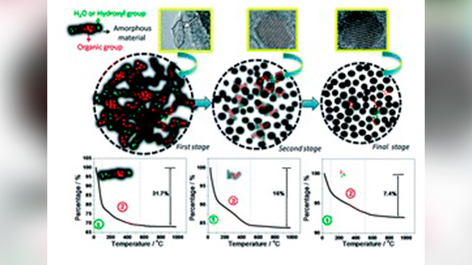
Abstract: We report on the synthesis and characterization of ZrO2 nanoparticles prepared from zirconium(IV) butoxide in the absence of base or acid mineraliser by an advanced oxidation processes (AOP) and subsequent hydrothermal treatment which involves mixing and dissolution of the precursor for different temperatures. The structure, morphology and properties of the nanoparticles were characterized using XRD, FE-SEM, TEM, HRTEM, Raman spectroscopy, FT-IR spectroscopy, BET measurements and TGA measurements. The structural analysis by XRD and Raman spectroscopy confirms the ZrO2 specimens synthesized at 100 °C for 1 hour are amorphous and those treated at 150 °C and 200 °C for 1 hour were crystalline. Structural analysis by Raman spectroscopy confirms tetragonal ZrO2 specimens obtained at temperatures higher than 100 °C as a majority phase. From FE-SEM images, the AOP/hydrothermal route mainly produced microspheres comprised of primary nanoparticles. HRTEM images of ZrO2 microspheres, after treatment at 100 °C, show the beginning of crystallization, with only a few clear lattice fringes dispersed in the specimens. TEM and HRTEM images of ZrO2, after treatment at 150 °C and 200 °C indicate that the microspheres are the aggregation of small nanoparticles with a size of approximately 5–8 nm, and FFT analysis confirms the high crystallinity of the specimens. TGA analyses show a distinctive behavior for each of the ZrO2 specimens, after treatment at 100 °C, 150 °C and 200 °C. FT-IR analysis of ZrO2 specimens after hydrothermal treatment revealed the presence of groups, such as –OH, –CH2, and –CH at the surface. In addition, FT-IR spectra show a decrease in the amount of functional groups attached to the surface of the nanoparticles when the reaction temperature is increased. This result was also confirmed by TGA analysis. The ZrO2 specimens prepared by the AOP/hydrothermal route exhibited a high surface area of 511–184 m2 g−1.


