Writers: Pellissari, Claudia Viviane Guimaraes and Pavarina, Ana Claudia and Bagnato, Vanderlei Salvador and Mima, Ewerton Garcia de Oliveira and Vergani, Carlos Eduardo and Jorge, Janaina Habib
Keywords: Cells; microorganisms; cytotoxicity
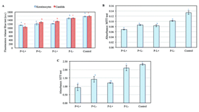
Abstract: This study assessed the cytotoxicity of antimicrobial Photodynamic Inactivation (aPDI), mediated by curcumin, using human keratinocytes co-cultured with Candida albicans. Cells and microorganisms were grown separately for 24 hours and then kept in contact for an additional 24 hours. After this period, aPDI was applied. The conditions tested were: P+L+ (experimental group aPDI); P−L+ (light emitting diode [LED] group); P+L− (curcumin group); and P−L− (cells in co-culture without curcumin nor LED). In addition, keratinocytes and C. albicans were grown separately, were not placed in the co-culture and did not receive aPDI (control group). Cell proliferation was assessed using Alamar Blue, MTT, XTT and CFU tests. Qualitative and quantitative analyses were performed. Analysis of variance (ANOVA) was applied to the survival percentages of cells compared to the control group (considered as 100% viability), complemented by multiple comparisons using Tukey’s test. A 5% significance level was adopted. The results of this study showed no interference in the metabolism of the cells in co-culture, since no differences were observed between the control group (cultured cells by themselves) and the P−L− group (co-culture cells without aPDI). The aPDI group reached the highest reduction (p = 0.009), which was equivalent to 1.7 log10 when compared to the control group. The P+L−, P−L+, P−L− and control groups were not statistically different (ρ > 0.05). aPDI inhibited the growth of keratinocytes and C. albicans in all tests, so the therapy was considered slightly (inhibition between 25 and 50% compared to the control group) to moderately (inhibition between 50 and 75% compared to the control group) cytotoxic.


