Writers: Leonardo Marques; Leandro A. Holgado; Rebeca D. Simões; João D. A. S. Pereira; Juliana F. Floriano; Lígia S. L. S. Mota; Carlos F. O. Graeff; Carlos J. L. Constantino; Miguel. A. Rodriguez-Perez; Mariza Matsumoto; and Angela Kinoshita
Keywords: biomaterial; piezoelectricity; tissue reaction; cytotoxicity; PVDF
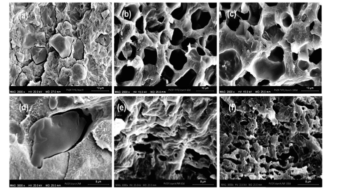
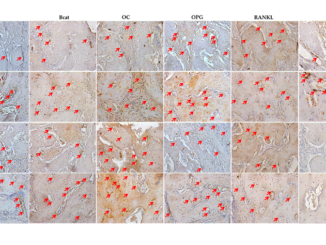
Abstract: Cytotoxicity and subcutaneous tissue reaction of innovative blends composed by polyvinylidene fluoride and polyvinylidene fluoride-trifluoroethylene associated with natural polymers (natural rubber and native starch) forming membranes were evaluated, aiming its applications associated with bone regeneration. Cytotoxicity was evaluated in mouse fibroblasts culture cells (NIH3T3) using trypan blue staining. Tissue response was in vivo evaluated by subcutaneous implantation of materials in rats, taking into account the presence of necrosis and connective tissue capsule around implanted materials after 7, 14, 21, 28, 35, 60, and 100 days of surgery. The pattern of inflammation was evaluated by histomorphometry of the inflammatory cells. Chemical and morphological changes of implanted materials after 60 and 100 days were evaluated by Fourier transform infrared (FTIR) absorption spectroscopy and scanning electron microscopy (SEM) images. Cytotoxicity tests indicated a good tolerance of the cells to the biomaterial. The in vivo tissue response of all studied materials showed normal inflammatory pattern, characterized by a reduction of polymorphonuclear leukocytes and an increase in mononuclear leukocytes over the time (p < 0.05 Kruskal–Wallis). On day 60, microscopic analysis showed regression of the chronic inflammatory process around all materials. FTIR showed no changes in chemical composition of materials due to implantation, whereas SEM demonstrated the delivery of starch in the medium. Therefore, the results of the tests performed in vitro and in vivo show that the innovative blends can further be used as biomaterials.


